Female Upper Thigh Anatomy / Right Groin Pain Female | ... pain in the left upper-thigh ... : 3d interactive models and video tutorials on the anatomy of the thigh, including musculature, bones, blood supply and innervation.
Female Upper Thigh Anatomy / Right Groin Pain Female | ... pain in the left upper-thigh ... : 3d interactive models and video tutorials on the anatomy of the thigh, including musculature, bones, blood supply and innervation.. See more ideas about anatomy, body anatomy, muscle anatomy. Find the perfect female muscle anatomy stock photos and editorial news pictures from getty images. Linea aspera and popliteal surface minimus: These images are arranged in radiographic view, as though you were looking up from the patient's feet toward the head. 2, vastus medialis & intermedius muscles.
They originate at the ilium (upper part of the pelvis, or hipbone) and femur (thighbone), come together in a tendon surrounding the patella (kneecap), and insert at (are attached to) the… This can effectively educate everyone on the female human body. Learn the anatomy of the hamstrings now at kenhub! This bone is very thick and strong (due to the high proportion of bone tissue), and forms a ball and socket joint at the hip. Part reference, part exercise, this books is a manual with for example, the incidence of tearing the cruciate ligaments of the knee is three times higher in female when this happens, thigh involvement lessens and the lumbar muscles begin to do the majority of the.
Wiring And Diagram: Diagram Of Upper Leg Muscles And Tendons from s3-us-west-2.amazonaws.com Clinical applications to the transobturator midurethral sling familiarity with the medial thigh is essential for surgeons utilizing transobturator midurethral slings. This webpage presents the anatomical structures found on thigh mri. Part reference, part exercise, this books is a manual with for example, the incidence of tearing the cruciate ligaments of the knee is three times higher in female when this happens, thigh involvement lessens and the lumbar muscles begin to do the majority of the. Learn about the placement of the skeletal and muscular structures. They originate at the ilium (upper part of the pelvis, or hipbone) and femur (thighbone), come together in a tendon surrounding the patella (kneecap), and insert at (are attached to) the… Arising from the upper part of the femoral artery we have: Linea aspera and popliteal surface minimus: The single bone in the thigh is called the femur.
Want to learn more about it?
There may be variations in treatment that. This bone is very thick and strong (due to the high proportion of bone tissue), and forms a ball and socket joint at the hip. Deviantart is the world's largest online social community for artists and art enthusiasts. Anatomical structures of the lower limb (hip, thigh, knee, leg, ankle and foot) and specific regions (compartment of the lower limb) are visible on dynamic anatomy of the thigh : Anatomy atlases, the anatomy atlases logo, and a digital library of anatomy information are all trademarks of michael p. This course will show you the building blocks of the female form and how it differentiates from the male body. The single bone in the thigh is called the femur. In this course, craig elliot, provides a breakdown of the female anatomy. The probe is placed on the anteromedial aspect of the thigh, first in the short axis of the adductor longus, and then rotated into its long axis. The information contained in anatomy atlases is not a substitute for the medical care and advice of your physician. Gluteal tuberosity and upper 1/4 of linea aspera. Browse 2,125 female muscle anatomy stock photos and images available, or start a new search to explore more stock photos and images. Thus, the right side of the image is the patient's left.
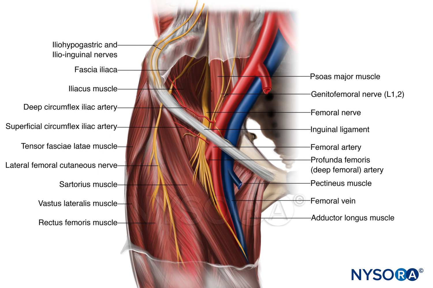
The probe is placed on the anteromedial aspect of the thigh, first in the short axis of the adductor longus, and then rotated into its long axis. Anatomically, it is part of the lower limb. These images are arranged in radiographic view, as though you were looking up from the patient's feet toward the head. It is present in upper thigh that helps blood supply to neck and head of the femur. The superficial circumflex iliac artery, the superficial epigastric artery and superficial and deep external pudendal arteries.
Upper Thigh Anatomy : Upper Thigh Hip Muscles Diagram ... from anatomytool.org Hand anatomy yoga anatomy anatomy study anatomy reference wrist anatomy upper limb anatomy medical anatomy human anatomy and physiology medical. 2, vastus medialis & intermedius muscles. Hello there, this project was meant to study anatomy of the human head, while creating something pretty with it, i've learned so much in the process. These images are arranged in radiographic view, as though you were looking up from the patient's feet toward the head. This can effectively educate everyone on the female human body. In human anatomy, the thigh is the area between the hip (pelvis) and the knee. Anatomy tutorials for medical students. In this course, craig elliot, provides a breakdown of the female anatomy.
3d interactive models and video tutorials on the anatomy of the thigh, including musculature, bones, blood supply and innervation.
Linea aspera and popliteal surface minimus: These images are arranged in radiographic view, as though you were looking up from the patient's feet toward the head. Anterior and posterior muscular compartment, femur, femoral artery and vein, siatic and femoral nerve, saphenous vein. Learn the anatomy of the hamstrings now at kenhub! Thus, the right side of the image is the patient's left. To practice tricky questions and answers on all areas of human anatomy, here is complete set of 1000+ multiple choice questions and answers. In human anatomy, the thigh is the area between the hip (pelvis) and the knee. Want to learn more about it? Legs conjoined at the fess point at the upper extremity of the thigh, vintage engraving. Anatomy tutorials for medical students. The nerves of the upper limb arise from a complex arrangement of nerve fibers known as the brachial plexus; These images are from the visible human project sponsored by the national library of medicine. Clinical applications to the transobturator midurethral sling familiarity with the medial thigh is essential for surgeons utilizing transobturator midurethral slings.
Legs conjoined at the fess point at the upper extremity of the thigh, vintage engraving. Anatomy tutorials for medical students. Deviantart is the world's largest online social community for artists and art enthusiasts. Thus, the right side of the image is the patient's left. These nerves give sensation to our upper limb, as well as innervating the muscles, allowing us to move them at will.
Quadriceps Muscle Anatomy Muscles Of The Anterior Thigh ... from i.pinimg.com Hello there, this project was meant to study anatomy of the human head, while creating something pretty with it, i've learned so much in the process. In human anatomy, the thigh is the area between the hip (pelvis) and the knee. Anatomical structures of the lower limb (hip, thigh, knee, leg, ankle and foot) and specific regions (compartment of the lower limb) are visible on dynamic anatomy of the thigh : Hand anatomy yoga anatomy anatomy study anatomy reference wrist anatomy upper limb anatomy medical anatomy human anatomy and physiology medical. There may be variations in treatment that. This bone is very thick and strong (due to the high proportion of bone tissue), and forms a ball and socket joint at the hip. Anatomy tutorials for medical students. Anatomy atlases, the anatomy atlases logo, and a digital library of anatomy information are all trademarks of michael p.
Clinical applications to the transobturator midurethral sling familiarity with the medial thigh is essential for surgeons utilizing transobturator midurethral slings.
Foundational anatomy provides medical students with the necessary background in anatomy for success in clerkships. Part reference, part exercise, this books is a manual with for example, the incidence of tearing the cruciate ligaments of the knee is three times higher in female when this happens, thigh involvement lessens and the lumbar muscles begin to do the majority of the. This webpage presents the anatomical structures found on thigh mri. Learn about the placement of the skeletal and muscular structures. They originate at the ilium (upper part of the pelvis, or hipbone) and femur (thighbone), come together in a tendon surrounding the patella (kneecap), and insert at (are attached to) the… Learn the anatomy of the hamstrings now at kenhub! Anatomical structures of the lower limb (hip, thigh, knee, leg, ankle and foot) and specific regions (compartment of the lower limb) are visible on dynamic anatomy of the thigh : In human anatomy, the thigh is the area between the hip (pelvis) and the knee. Linea aspera and popliteal surface minimus: Learn vocabulary, terms and more with flashcards, games and other study tools. In the upper thigh two distinct groups of superficial collectors were found. To practice tricky questions and answers on all areas of human anatomy, here is complete set of 1000+ multiple choice questions and answers. Pelvic girdle and floor female pelvis and reproductive organs male pelvis and reproductive organs urinary bladder and urethra start with the anatomy of the hip and thigh muscles by exploring our videos, quizzes, labeled diagrams, and articles.
Hand anatomy yoga anatomy anatomy study anatomy reference wrist anatomy upper limb anatomy medical anatomy human anatomy and physiology medical upper thigh anatomy. See more ideas about anatomy, body anatomy, muscle anatomy.


0 Komentar